Precision at scale: injection landmarking for general practice
Amy Shirtliff BA (Hons) MSc RGN, Nurse Practitioner & Writer
Practice Nurse 2026;56(5):21-24
A misplaced deltoid intramuscular injection can result in severe, persistent shoulder pain and a marked reduction in range of motion, adverse effects that are entirely preventable with correct landmarking
General practice nurses (GPNs) give hundreds of deltoid intramuscular (IM) injections under significant time pressure, and autumn’s flu season brings the most intense operational demands of the year to the surgery.1 High-volume repetition carries an inherent risk of automation, and when a procedure is performed hundreds of times, the hand starts moving to inject before the eye has checked the landmark.2
This article focuses on the exact anatomical landmarking needed to isolate the safe zone, building on general administration guidelines and focusing on landmarking as a specific skill. By concentrating on the deltoid during peak flu season, and then extending to other critical sites such as the ventrogluteal region, GPNs can maintain accurate technique even in the busiest clinics.3
Landmarking has two components:
- Surface location: positioning the needle on the correct skin coordinate, to avoid the underlying nerves, bursae, and joint capsules.
- Tissue depth: ensuring the needle passes through the subcutaneous fat and into the target muscle belly itself.
The deltoid: a precise target
The safe zone for a deltoid injection is significantly smaller than the visible muscular contour of the upper arm, and it is easy to misjudge and assume the safe zone is larger than it is. Relying on visual estimation alone risks mistargeting the vulnerable structural boundaries that sit just outside it.
Anatomical boundaries:
- Superior boundary: the acromion process: the hard, bony ridge at the top of the shoulder.
- Inferior boundary: the deltoid tuberosity on the mid-shaft of the humerus.
- Internal landmarks: the subdeltoid/subacromial bursa and the axillary nerve, which courses horizontally around the humerus roughly 3 to 5 cm below the acromion.
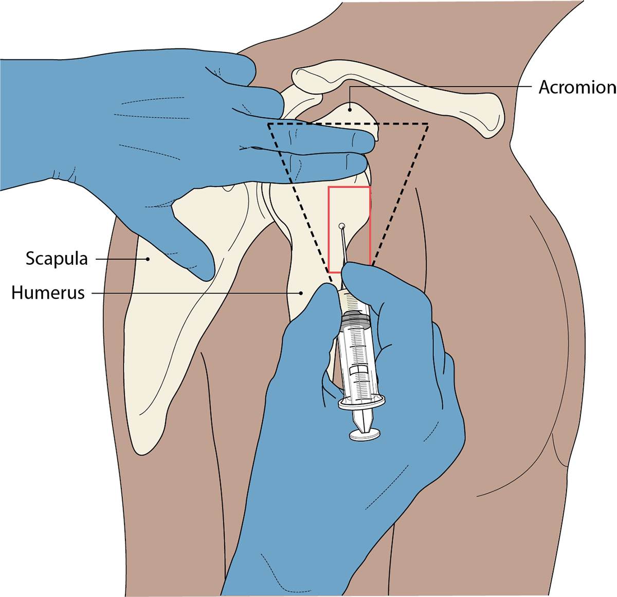
To locate the safe zone, start at the top of the shoulder girdle and palpate the lower edge of the acromion process. Drop 2.5 to 5 cm (2–3 finger-widths) straight down along the lateral aspect of the arm. The target zone sits in the dense, central bulk of the muscle belly, well above the axillary fold line (Figure 1).4
Operational causes of injection site misplacement
The anatomical landmarks themselves never move, but the variables and everyday conditions of a real clinic make good technique harder to maintain:
- Nurse-patient height discrepancy. If a patient stays standing, or sits high on an examination couch, the nurse is forced to reach up, and that distorts the perpendicular 90-degree entry angle. Slanting the needle upward sends it through subcutaneous fat at an oblique angle rather than penetrating cleanly into the muscle belly. Seating the patient brings the shoulder down, ensuring a true 90-degree entry straight into the central muscle mass.5
- Inadequate clothing exposure. When a patient pulls a shirt collar laterally rather than exposing the entire arm, only the acromial cap and the uppermost deltoid are visible, and injecting into that exposed skin forces placement into the high-risk subdeltoid zone.
- Constricting sleeve bands. A tight sleeve rolled up the upper arm constricts the upper arm. It prevents clear top-down palpation of the acromion, distorts the tissue layers, and stops the muscle hanging in a relaxed state.
- Muscle tension and shrugging. A tensed, unsupported arm elevates the shoulder girdle, shifting the relative surface landmarks upward and obscuring the tissue boundaries beneath.
SIRVA
SIRVA (Shoulder Injury Related to Vaccine Administration) is thought to be a key clinical consequence of an injection placed too high on the arm, occurring when vaccine antigen and adjuvant are inadvertently deposited into the subdeltoid bursa, the synovial capsule, or the rotator cuff tendons, rather than into the deltoid muscle belly.
Pathophysiology: Deltoid muscle tissue tolerates immune activation with minimal localised damage, whereas the avascular, enclosed environment of the subdeltoid bursa does not. Exposure to adjuvanted vaccine at this site is thought to trigger a prolonged inflammatory response, which may manifest as subdeltoid bursitis, adhesive capsulitis (frozen shoulder), tendonitis or rotator cuff tears.
Clinical presentation: Ordinary post-vaccination reactogenicity usually settles within 24 to 48 hours, but SIRVA tends not to. It typically presents as severe, persistent shoulder pain and a marked reduction in range of motion, specifically during active and passive abduction and external rotation. Symptoms characteristically begin within 48 hours of administration, and often fail to respond to standard first-line oral analgesia or NSAIDs. SIRVA is widely regarded as iatrogenic and entirely preventable, stemming from flawed surface landmarking rather than from vaccine toxicity or contamination.6
Depth: reaching the muscle
Surface placement alone is not enough. The needle also has to reach the intramuscular layer for the antigen to be delivered as intended. In the deltoid, depth errors can go either way; too deep, striking bone, or too shallow, but it is shallow deposition into subcutaneous fat that most often undermines the vaccine. Delivering an intramuscular formulation into subcutaneous tissue can cause two problems. The first is elevated local reactogenicity; aluminium-adjuvanted vaccines in particular left sitting in fat layers can elicit localised tissue necrosis, persistent subcutaneous nodules, and sterile abscesses. The second is reduced immunogenicity, because subcutaneous fat lacks the dense vascularity and the antigen-presenting cell networks required for optimal vaccine uptake.7
- Managing needle selection. Choosing the right needle length and gauge is an integral component of tissue-depth landmarking:
- Standard adult (standard mass): a 25mm needle (commonly 23G blue or 25G orange) provides sufficient penetration into the central deltoid belly for most adults.
- High BMI / large muscle mass: the subcutaneous adipose tissue over the deltoid expands significantly, and a standard 25mm needle risks terminating in that subcutaneous layer. A 38 mm needle (21G green, or a long 23G blue) may be required to reliably penetrate muscle belly tissue.3
- Frail/low BMI adults: The Green Book does not provide guidance pertaining specifically to adults with low BMI, or noticeably minimal subcutaneous fat or low deltoid muscle mass.3 A 25mm needle remains standard; however, it is important to note that an emerging body of evidence suggests the greater risk in frail adults with deltoid sarcopenia is overpenetration. With the muscle reduced to a thin layer within fat, a needle can pass through it into the underlying neurovascular bundle. In these patients the muscle can be gently bunched rather than stretched flat; evidence suggests stretching is strongly associated with injecting into that bundle.8
- Health equity in administration: Defaulting to a standard 25mm needle without assessing tissue depth may disproportionately affect patients with a higher body mass. Subcutaneous depth varies between individuals and is associated with factors including BMI, sex, and age. A one-size-fits-all approach to needle length can therefore increase the risk of subcutaneous deposition in some patients. Individual assessment supports consistent delivery into muscle.9
Beyond the deltoid: general practice pitfalls
Flu season keeps our attention on the deltoid, but several other routine GPN procedures carry critical landmarking risks of their own.
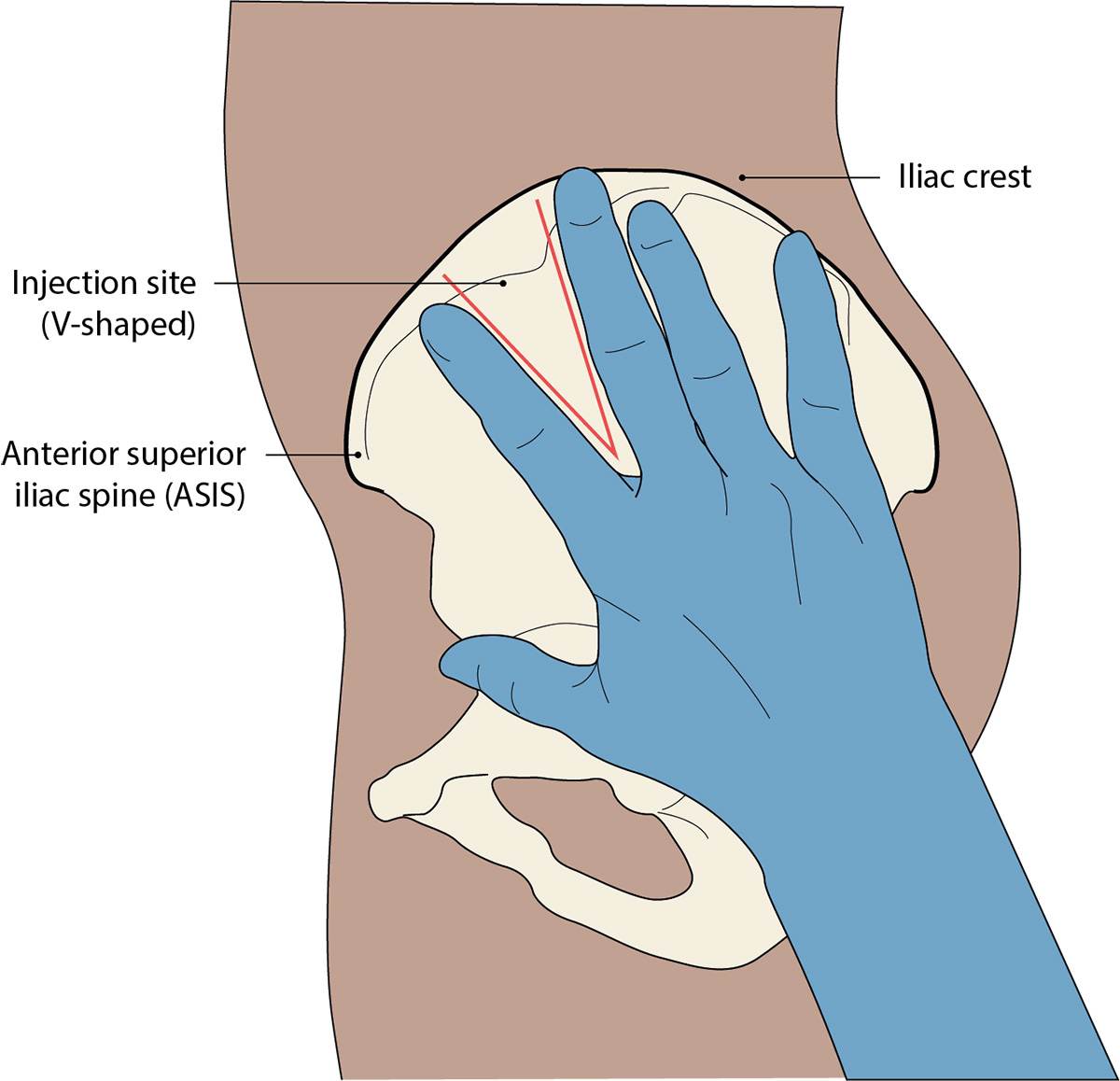
Ventrogluteal versus dorsogluteal (contraceptive depot injections): The dorsogluteal site, the upper outer quadrant of the buttock, remains an ingrained habit in general practice, yet it poses a well-documented risk of sciatic nerve damage, as well as inadvertent intravascular or deep subcutaneous deposition. The ventrogluteal site is the safer choice (Figure 2), by avoiding the sciatic nerve entirely, sitting clear of the major gluteal vessels, and is covered by a significantly thinner layer of subcutaneous fat.
The ventrogluteal landmark technique:
- Position the palm of your hand over the patient's greater trochanter (right hand for the left hip, left hand for the right hip).
- Point your index finger toward the anterior superior iliac spine (ASIS).
- Spread your middle finger posteriorly along the iliac crest to form a V shape.
- Insert the needle at 90 degrees into the centre of the V.10
Vastus lateralis (paediatric immunisations): For infants under one year, the anterolateral thigh, the vastus lateralis, is the primary target site, offering a large muscle mass that is clear of the major nerves and vessels. Aim for the middle third of the muscle on the anterolateral aspect of the thigh. For the tissue technique, stabilise the muscle by holding the skin taut.3
Co-administering multiple vaccines (flu, COVID-19 and RSV): Autumn GPN workloads increasingly involve giving more than one vaccine (influenza, a COVID-19 booster, or RSV) in a single consultation. Where feasible, administer the vaccines into opposite arms, which enables precise monitoring of the localised reactogenicity of each. When the same arm has to be used, separate the injection sites by at least 2.5 cm along the longitudinal axis of the deltoid belly, to prevent overlapping local inflammatory responses. Record explicitly which vaccine was administered into which site and arm in the patient's record, to maintain the safety audit trail.3
Limb avoidance: Site selection always requires evaluating the patient's full medical background. Following breast cancer treatment involving the axilla, there is no consistent evidence that injection on the treated side increases lymphoedema risk. Where alternatives are readily available, they may be preferred, but NICE guidance updated in 2025 recommends as a shared decision based on patient preference, clinical judgement and practical alternatives.11In other cases, other limbs are avoided for different reasons. Steer clear of an arm with a functioning arteriovenous fistula or graft used for haemodialysis, both to protect the access and to avoid introducing infection.12 An arm with significantly impaired sensation, movement or circulation, for instance on the affected side after a stroke, is also best avoided where an alternative exists, since the patient may not reliably feel or report a local complication.13
Quick fixes
High-throughput flu clinics create environmental pressures that undermine landmarking accuracy, and protecting patient safety requires fast, systematic fixes for each predictable failure:
- Eyeball landmarking: Time pressure might tempt us to skip manual palpation altogether. The quick fix, ‘palpate then drop’, takes seconds. Palpate the acromion edge at the top of the shoulder, drop 2.5–5 cm (2–3 finger-widths) down, and only then target the central muscle belly.3
- Upward needle trajectory: A patient who is standing, or seated high, draws the nurse's reach upward into the subdeltoid bursa zone. The quick fix is mandatory seating and ensures all patients sit in a standard chair, with the arm resting unsupported in their lap.5
- Sleeve restriction: Short or tight sleeves roll up into a constricting band. The quick fix, the “arm-out rule”, is that if a rolled sleeve restricts a clear view of the acromion and the full deltoid belly, the arm comes fully out of the garment. Stating a preference for sleeveless garments in vaccine invitation letters would facilitate this.6
- Default needle selection: A 25mm needle isn’t right for everyone. The quick fix, a visual depth check for every patient, ensures arm size is assessed before reaching for a needle.3
In the background to a safe and unhurried clinic is an efficient patient flow through the surgery. A one-way, production-line system works well: patients are marked as arrived and directed in at one point, move on to a vaccinating station where consent and eligibility are checked and the injection is given, and leave past a clearly signposted point displaying post-vaccination advice. Keeping people moving in a single direction avoids bottlenecks and lets the vaccinator concentrate on technique rather than logistics.14
Conclusion
Injection technique is not a fixed skill. Even procedures done thousands of times deserve periodic technique re-examination. The deltoid safe zone in particular is physically smaller than the visible arm contour, and high-volume clinics only increase the risk of misplacement.4The safeguards are quick and simple: manual, top-down palpation for the surface, and a systematic depth assessment for the tissue.3 By relying on these two habits, clinicians can consistently deliver safe, effective injections even in the most fast-paced clinic settings.
KEY POINTS |
|
References
1. The Queen’s Institute of Community Nursing. General Practice Nursing Today; 2026. https://qicn.org.uk/wp-content/uploads/2026/05/General-Practice-Nursing-Today-2026.pdf
2. Greenway K. Rituals in nursing: intramuscular injections. J Clin Nurs 2014;23(23-24):3583-8
3. UK Health Security Agency. Immunisation procedures: the green book, chapter 4. 2013. https://assets.publishing.service.gov.uk/media/5a7afc62e5274a34770e88e5/Green-Book-Chapter-4.pdf
4. Charmode S, Sharma S, Kushwaha S, et al. Deltoid Intramuscular Injections: A Systematic Review of Underlying Neurovascular Structures to the Muscle and Proposing a Relatively Safer Site. Cureus 2022;14(4):e24172
5.UK Health Security Agency. National minimum standards and core curriculum for vaccination training; 2025. https://assets.publishing.service.gov.uk/media/6855b286b46781eacfd71dc9/UKHSA_National_Minimum_Standards_for_immunisation_training_2025.pdf
6. Bancsi A, Houle S, Grindrod K. Getting it in the right spot: Shoulder injury related to vaccine administration (SIRVA) and other injection site events. Can Pharm J 2018;151(5):295–299
7. Zuckerman J. The importance of injecting vaccines into muscle: Different patients need different needle sizes. BMJ 2000;321(7271):1237–1238
8. Davidson K, Bertram J. Best practice for deltoid intramuscular injections in older adults: Study in cadavers. J Nurs Educat Pract2019;9(9):92-97
9. Sebro R. Statistical estimation of deltoid subcutaneous fat pad thickness: implications for needle length for vaccination.Sci Rep 2022;12(1):1069
10. Gutierrez J, Munakomi S. Intramuscular Injection. Stat Pearls 2026 Jan-. https://www.ncbi.nlm.nih.gov/books/NBK556121/
11. NICE NG101. Early and locally advanced breast cancer: diagnosis and management; 2025. https://www.nice.org.uk/guidance/ng101
12. Government of Canada. Vaccine administration practices: Canadian Immunization Guide; 2026. https://www.canada.ca/en/public-health/services/publications/healthy-living/canadian-immunization-guide-part-1-key-immunization-information/page-8-vaccine-administration-practices.html
13. Cambridge University Hospitals NHS Foundation Trust. Post-stroke sensory deficits and re-education. https://www.cuh.nhs.uk/patient-information/post-stroke-sensory-deficits-and-re-education/
14. Shirtliff A. Preparing for the flu season during the COVID-19 outbreak. Practice Nurse 2020;50(7):10-15
Related articles
View all Articles